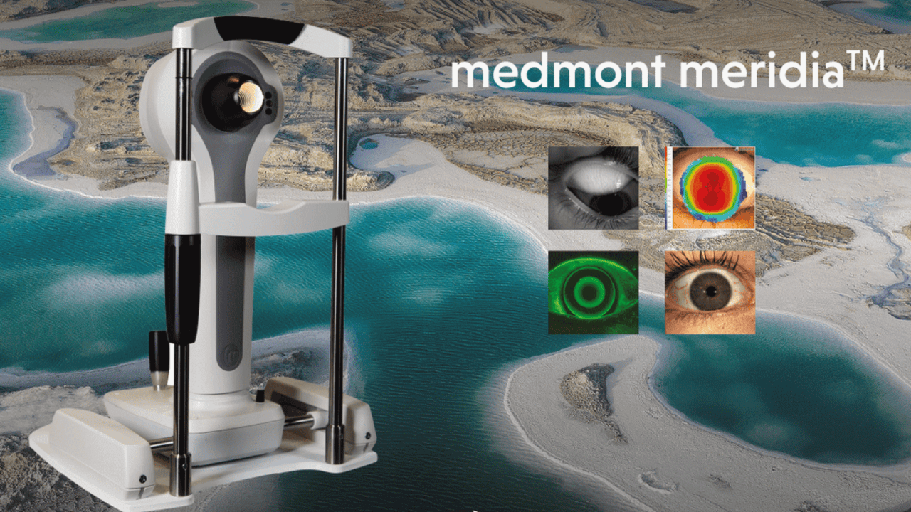
The Medmont Meridia™ creates precise topographies of the cornea from limbus to limbus. With the camera's high-resolution color images and many other functions, the optimal contact lens can be fitted. Indispensable for orthokeratology, scleral lenses or other special lenses.
The Medmont Meridia™ Advanced Corneal Topographer – Empowering Healthy Sight

The Medmont Meridia™ Advanced Corneal Topographer has outstanding and versatile image and video analysis especially for the dry eye. It creates large and precise topographies of the cornea from limbus to limbus. With the high-resolution color images of the camera and many other functions, the optimal contact lens can be fitted. Indispensable for orthokeratology, scleral lenses or other special lenses.
Fast adopted by contact lens professionals around the world, the Medmont Meridia™ builds on the Medmont E300’s goldstandard topography with full-colour maps, an ultra-wide field of view and unmatched corneal coverage.
The Meridia™ Pro introduces many new modes for anterior eye imaging—dry eye (DED) analysis and reporting, tear film analysis, meibography and more. It’s everything you need for patient and practice success in one compact instrument.
Small cone design
The shorter working distance of the Meridia™ projects more rings onto the corneathan a large cone, offering a much larger capture field and a higher concentrationof measured points to more accurately determine corneal shape.
Wide field-of-view
The Meridia™'s 16mm field of view enables quick, easy visualisation of the eye. Measure the full HVID, pick up anomalies without moving to anterior analysis—and more. Reclaim time in your busy schedule by saving steps in your workflow.
Ultra-wide single capture

Generate up to 11mm+ of real corneal data in a single capture—without extrapolation. Fast-track excellent contact lens outcomes with all the information you need at a glance, so you can see more patients and generate more revenue.
Composite topography
Use composite imaging to extend real data coverage to the edge of the sclera. It captures five images in different gaze directions and stitches them into one so you can measure and map limbus-to-limbus—invaluable for large lens design.
Effortless auto capture The Meridia™ initiates capture when you reach minimum alignment. Your job is to center the green cross hair and ensure an even tear film, guaranteeing distortion-free rings and precise data capture. Then, choose the most optimal map. It’s simple.
Full colour topography
See the anterior eye itself in brilliant HD colour during corneal topography. This lifelike view of the cornea and sclera lets you better observe subtle details and identify conditions that aren’t immediately apparent with black and white imaging.
Digital zoom
Use the Meridia™'s 2-10x digital zoom to zone in on regions of interest. With the Meridia™ Pro, you can save zoomed areas in all anterior capture modes. Use the HD images for documentation and to demonstrate pathology to your patients.
Quick keys
With the Meridia™'s convenient new quick keys, you can stay stationed at your instrument instead of turning to your keyboard or mouse to capture images and video. It all adds up to a faster, smoother workflow.
Contact lens simulator
Build custom lenses by determining path and custom parameters for each patient, and assess how a lens will look on the eye. This increases your first-fit success for less trial lenses and exchanges. Increase your efficiency and proficiency, and see more patients.
Anterior imaging and video
Capture and document high-quality colour images and video for well-informed clinical decisions, patient education and high confidence in your treatment plans. Easily export them to colleagues for collaboration or to labs for contact lens design.
Single-operator meibography

Screen, track and document Meibomian Gland Disorder with ease. The Meridia™’s short working distance means you can hold the patient’s lids and capture images at the same time. No assistant required.
Fluorescein review
See and document contact lens fits and corneal surface health with ultra-clear Fluorescein imaging and video. It activates cobalt light to highlight any conjunctival or corneal staining, as well as aiding tear film assessment.
Tear Meniscus Height (TMH) measurements
Quickly gain a quantitative tear volume measurement for an important piece of the DED subtyping puzzle—with minimal effort. Use the TMH annotation tool on any anterior image or topography capture for one less step in your workflow.
Tear film analysis mode

The Meridia™ offers a non-invasive tear break up time test (‘NIBUT’) that measures in seconds to 1 decimal place—without sodium fluorescein dye. You can also map tear film surface quality (TFSQ) to identify and locate unstable tear film.
Universal dry eye grading

Ensure image gradings are reliably universal throughout your practice with standardised dry eye scales (Efron, BHVI, Meiboscale). Use them to educate patients on their level of disease and monitor outcomes to inform treatment plans.
Comprehensive dry eye reports

Quickly pull your dry eye workflow tests and images into a visual report. Use it to present findings to your patient to help them understand your diagnosis and recommendations. Generate a new report at each visit to track changes over time.
The Medmont Meridia™ thus offers versatile image and video analyses specifically for the dry eye in addition to the large-area topographical recording of the cornea from limbus to
limbus. This provides the user with a comprehensive modern corneal topographer with further outstanding and versatile functions for the practice.


